Catalog Number: HK-CAF23
Product Format: Frozen Vial
Cell Number: 1,000,000 cells/vial
Suggested Medium: CAF Growth Medium (CAF01)
General Information
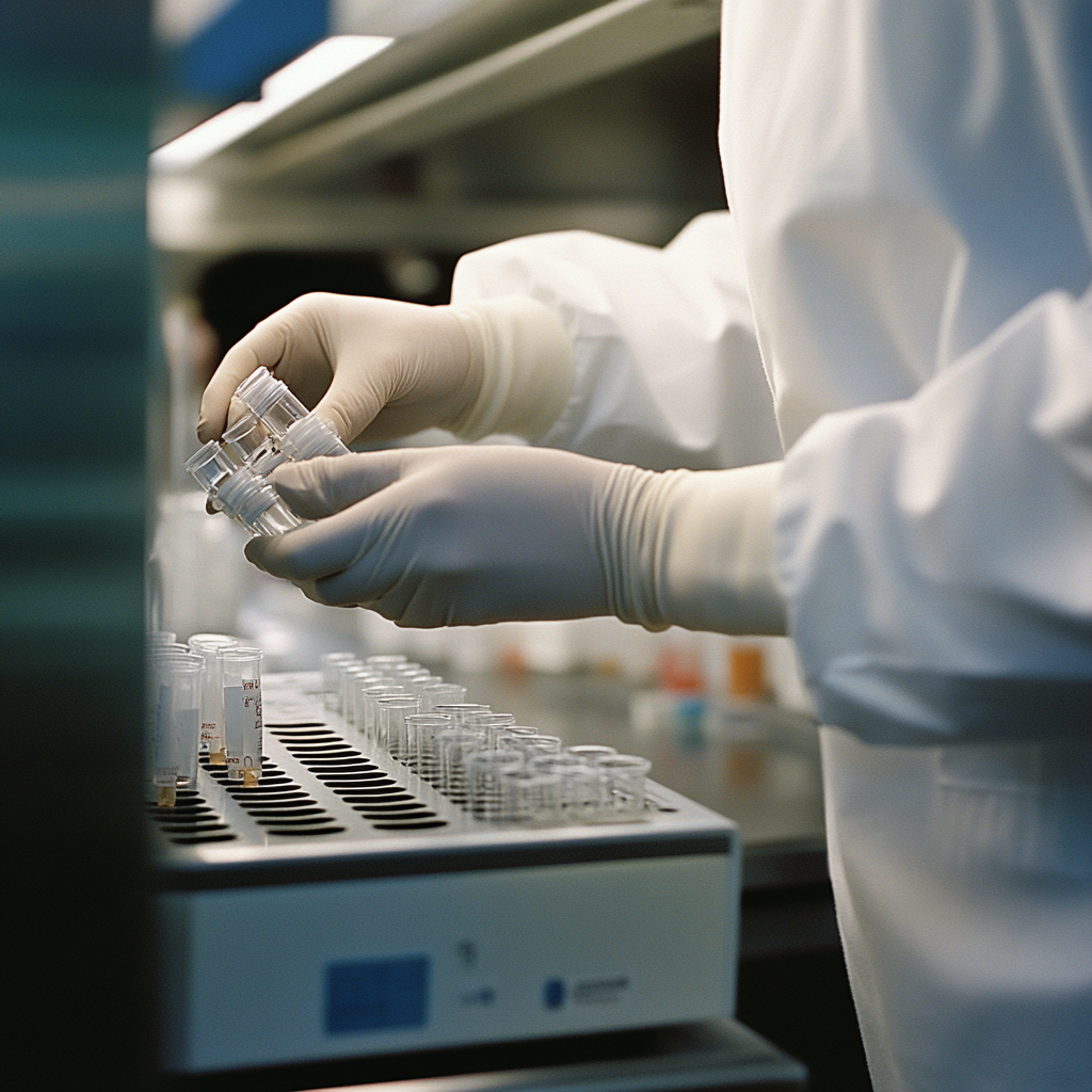
Human Kidney CAF is isolated from human kidney tumor tissue. Cells are grown in T75 tissue culture flasks pre-coated with AlphaBioCoat solution for 30 minutes and incubated in CAF Growth Medium for 3–7 days. Cultures are then expanded. Prior to shipping, cells at passage 1 are detached from flasks and immediately cryo-preserved in vials. Each vial contains at least 1,000,000 cells per ml.
Product Testing
Human Kidney CAF is tested for negative expression of von Willebrand Factor Expression/Factor VIII, cytokeratin 18, and alpha-smooth muscle actin. Cells are negative for bacteria, yeast, fungi, and mycoplasma.
Cells express CAF markers FAP, PDGFR, Vimentin, PDPN, and CD70.
Cells can be expanded for 3–5 passages at a split ratio of 1:2 or 1:3.
Laboratory Applications
Human Kidney CAF can be used for the assay of cell-cell interaction, adhesion, PCR, Western blot, immunoprecipitation, immunofluorescent flow cytometry, or generating cell derivatives for desired research applications.
Shipping
Frozen Vials on Dry Ice
To ensure the highest level of viability, thaw the vial and initiate the culture as soon as possible upon receipt. If upon arrival, continued storage of the frozen culture is necessary, it should be stored in the liquid nitrogen vapor phase and not at -70°C. Storage at -70°C will result in loss of viability.
Thaw the vial by gentle agitation in a 37°C water bath. To reduce the possibility of contamination, keep the O-ring and cap out of the water. Thawing should be rapid (approximately 2 minutes).
Post-Thaw Handling Procedure
-
Remove the vial from the water bath as soon as the contents are thawed and decontaminate by dipping in or spraying with 70% ethanol. All operations from this point on should be carried out under strict aseptic conditions.
-
It is recommended that the cryoprotective agent be removed immediately. Centrifuge the cell suspension at approximately 125 × g for 5 to 10 minutes. Discard the supernatant and resuspend the cell pellet in an appropriate amount of fresh growth medium.
-
Add 6.0 to 8.0 mL of AlphaBioCoat to the T-Flask for 15 minutes. Aspirate the solution, rinse with 8 mL of 1× PBS, and discard the rinse. Transfer cells to an appropriate size T-Flask.
-
Avoid excessive alkalinity during recovery. Place the culture vessel with growth medium into the incubator for at least 15 minutes before adding thawed cells, allowing the medium to reach normal pH (7.0–7.6).
-
Incubate the culture at 37°C in a suitable incubator. A 5% CO₂ in air atmosphere is recommended if using the medium described.
Subculturing Procedure
Volumes are for a 75 cm² flask; adjust proportionally for other vessel sizes. T-75 flasks are recommended.
Note: To avoid clumping, do not agitate cells by hitting or shaking the flask during detachment. Cells that are difficult to detach may be placed at 37°C to facilitate dispersal.
-
Remove and discard the culture medium. Briefly rinse the cell layer with 1× PBS to eliminate serum traces.
-
Add 2.0 to 3.0 mL of Cell Detachment Solution and monitor under an inverted microscope until cells detach (typically within 5–15 minutes).
-
Add 6.0 to 8.0 mL of AlphaBioCoat to the T-Flask for 15 minutes. Aspirate and rinse with 8 mL of 1× PBS. Discard the rinse.
-
Add 6.0 to 8.0 mL of complete growth medium and gently aspirate cells.
-
Dispense appropriate aliquots into new culture vessels.
-
Incubate at 37°C.
Reagents for Cryopreservation
Complete growth medium supplemented with 5% (v/v) DMSO.
Catalog #: CGM5